
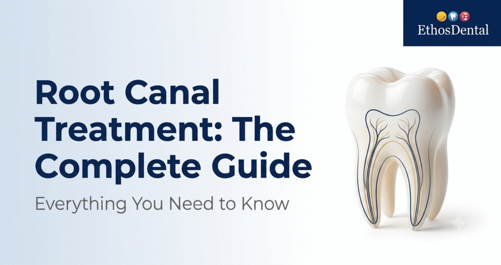
Root Canal Treatment: The Complete Guide Everything You Need to Know
By the clinical team at Ethos Dental • Updated 2026
Root canal treatment is one of the most misunderstood procedures in dentistry. Feared by patients, underestimated in complexity, and undervalued in outcome. This guide covers everything: the anatomy, the diagnosis, the step-by-step procedure, the materials, the technology, the success rates, and what every patient needs to know before making a decision about their tooth.
1. What Is Root Canal Treatment?
Root canal treatment (RCT), also called endodontic therapy, removes infected or inflamed pulp tissue from inside a tooth. The pulp the soft tissue containing nerves, blood vessels, and connective tissue can become infected through decay, cracks, trauma, or gum disease. Once infected, the pulp cannot heal on its own.
After removal, the canal system is cleaned, shaped, disinfected, and sealed with a biocompatible material. The tooth is then restored with a crown. The natural tooth remains in place, fully functional, for decades.
2. Tooth Anatomy — Understanding the Pulp
- Enamel — outermost hard layer protecting the crown
- Dentin — second layer with microscopic tubules leading to the pulp
- Pulp — innermost soft tissue — nerves, arteries, veins, lymphatics
- Cementum — covers the root, anchors the tooth to bone via the PDL
- Periodontal Ligament (PDL) — fibrous tissue connecting root to alveolar bone
Canals per tooth type:
- Incisors & canines — 1 canal
- Premolars — 1–2 canals
- Molars — 3–4 canals (upper first molars often have a hidden MB2 canal)
Even after pulp removal, the tooth survives because the surrounding periodontal tissues continue to nourish it. A root canal-treated tooth, properly restored, can last a lifetime.
How Infection Reaches the Pulp
- Deep caries — cavity progresses through enamel and dentin into pulp
- Cracked tooth — exposes pulp to oral bacteria
- Dental trauma — disrupts blood supply even without a visible fracture
- Repeated restorations — cumulative stress over time
- Periodontal disease — bacteria track up the root surface
3. Symptoms & Diagnosis
Primary Symptoms
- Severe, spontaneous toothache especially at night
- Prolonged sensitivity to hot or cold (>30 seconds after stimulus removed)
- Pain on biting or chewing
- Tooth darkening or discolouration
- Swelling of gum, face, or jaw
- Sinus tract (pimple-like bump on gum) sign of chronic abscess
- Persistent bad taste or odour
Asymptomatic infections are common. A dark shadow at the root tip on X-ray (periapical radiolucency) can indicate bone resorption from a silent chronic infection. Regular dental X-rays catch these before they become emergencies.
Pulp Diagnosis Classification (AAE System)
- Normal pulp — no treatment needed
- Reversible pulpitis — treat the cause; pulp can recover
- Irreversible pulpitis — RCT required
- Pulp necrosis — pulp is dead; RCT or extraction
- Previously treated — may need retreatment
Diagnostic Tests We Use
- Thermal testing — cold/heat to assess pulp vitality
- Electric pulp test (EPT) — low current to determine nerve response
- Percussion test — tapping to detect periapical inflammation
- Periapical X-ray — gold standard for periapical pathology
- CBCT 3D imaging — complex anatomy, missed canals, surgical planning
| Emergency Warning Signs — Seek Care Immediately ⚠ Facial swelling spreading toward the eye or neck ⚠ Difficulty breathing or swallowing ⚠ High fever with dental pain ⚠ Rapidly expanding jaw swelling |
4. The Root Canal Procedure Step by Step
1. Local Anaesthesia
2% Lignocaine with 1:80,000 adrenaline is the standard. For acute infections or anxious patients, supplemental techniques (intraligamentary or intrapulpal injection) ensure complete numbness.
2. Rubber Dam Isolation
Non-negotiable. Isolates the tooth from saliva and oral bacteria. Improves outcomes. Prevents accidental ingestion of irrigants or instruments.
3. Access Cavity Preparation
A precise opening through the crown into the pulp chamber. Conservative “micro-endodontic” access preserves maximum tooth structure.
4. Working Length Determination
Electronic apex locator (EAL) + confirmatory X-ray. Accurate to the apical constriction — prevents over- or under-instrumentation.
5. Canal Shaping — Rotary NiTi
ProTaper Gold, WaveOne Gold, or Reciproc Blue systems shape canals to the correct taper with minimal chair time. Heat-treated NiTi alloys flex in curved canals without fracturing.
6. Irrigation & Disinfection
Multi-irrigant protocol: • NaOCl 2.5–5.25% dissolves organic tissue, kills bacteria • EDTA 17% removes smear layer, opens dentinal tubules • CHX 2% residual antibacterial effect • Passive Ultrasonic Irrigation (PUI) drives irrigants into lateral canals and fins
7. Canal Drying
Sterile paper points. A moist canal compromises the seal.
8. Obturation
Canals filled with gutta-percha and bioceramic sealer. Warm vertical compaction or single-cone technique. Goal: 3D hermetic seal.
9. Coronal Restoration
Core build-up and crown. All posterior RCT teeth require a crown — this is the most important determinant of long-term success.
5. Single-Visit vs Multi-Visit Root Canal
| Entity | Single-visit RCT | Multi-visit RCT | Extraction |
| Sessions | 1 visit | 2–3 visits | 1 visit |
| Saves tooth | ✔ Yes | ✔ Yes | ✘ No |
| Post-op comfort | High | Moderate | Variable |
| Long-term value | Excellent | Excellent | Costly replacement |
| Best for | Uncomplicated cases | Acute infection / complex | Non-restorable tooth |
Current evidence: single-visit and multi-visit RCT have equivalent long-term success rates. Single-visit is preferred for uncomplicated cases. Multi-visit is clinically appropriate for acute apical abscess, complex anatomy, or when MTA is used for perforation repair.
6. Advanced Technology in Endodontics
Rotary NiTi Instrumentation
Nickel-Titanium rotary systems (ProTaper Gold, WaveOne Gold, Reciproc Blue) shape canals in minutes with superior flexibility in curved roots. Heat treatment (Gold or Blue wire) dramatically reduces fracture risk.
Electronic Apex Locator
Precisely locates the apical constriction electronically. Reduces radiation exposure by minimising confirmatory X-rays.
Dental Microscope / Magnification Loupes
Missed canals are the leading cause of RCT failure. The MB2 canal of upper first molars is missed in up to 40% of cases without magnification. Microscopic endodontics identifies all canals, micro-cracks, and calcified orifices.
CBCT 3D Imaging
Essential for retreatment, unusual anatomy (C-shaped canals, dens invaginatus), assessing the true extent of periapical lesions, and surgical planning.
Passive Ultrasonic Irrigation (PUI)
Acoustic streaming and cavitation drive irrigants into lateral canals, anastomoses, and fins inaccessible to instruments. Significantly improves disinfection in complex root systems.
Bioceramic Sealers
AH Plus Bioceramic, BioRoot RCS, TotalFill BC Sealer chemically bond to dentin, expand slightly on setting (eliminating microleakage), and are highly biocompatible. A generational improvement over traditional ZOE sealers.
7. Endodontic Materials
| Material | Properties | Best Used For |
| Gutta-Percha | Biocompatible, radiopaque, thermoplastic | Standard obturation — gold standard |
| Bioceramic Sealers | Calcium silicate-based, antibacterial | Superior seal, anti-resorptive |
| MTA | Biocompatible, moisture-tolerant | Perforation repair, apexification |
| Calcium Hydroxide | Antimicrobial, alkaline | Inter-appointment dressing |
| EDTA 17% | Chelating agent | Smear layer removal |
| NaOCl 2.5–5.25% | Dissolves organic tissue, antibacterial | Primary irrigant |
8. Types of Endodontic Procedures
Vital Pulp Therapy (Pulp Capping / Pulpotomy)
When pulp is still vital but exposed or inflamed, we may preserve vitality before committing to full RCT. Direct pulp cap with MTA or Biodentine. Pulpotomy removes coronal pulp only. Success rates with bioceramic materials exceed 90% at 5 years.
Conventional Root Canal Treatment (Pulpectomy)
Complete pulp removal from crown and roots. Standard procedure for irreversible pulpitis and pulp necrosis in permanent teeth.
Endodontic Retreatment
Removal of existing gutta-percha and sealer, re-instrumentation, identification of missed canals, and re-obturation. Indicated when a previously treated tooth develops new symptoms or radiographic failure.
Endodontic Microsurgery (Apicoectomy)
Surgical removal of the root tip and periapical lesion through a gum incision. Retrograde filling with MTA from the surgical approach. Success rates >90% with bioceramic materials and magnification.
Regenerative Endodontics
For immature permanent teeth with open apices. Disinfection followed by induction of bleeding to create a blood clot scaffold. MTA or Biodentine seals the orifice. Goal: continued root development and potential pulp revascularisation — a living tooth.
MTA Apexification
For immature teeth where regeneration is not possible. MTA apical plug creates an artificial apical stop in a single visit, allowing immediate obturation.
9. Complications & Management
Instrument Separation
NiTi file fracture within the canal. Prevention: single-use files, proper glide path, avoiding torque overload. Management: bypass, ultrasonic retrieval, or leaving in situ if prognosis unaffected.
Ledge Formation
Artificial blockage from improper technique. Prevented by establishing a glide path with hand files (K-files #08–15) before rotary instrumentation.
Perforation
Accidental communication between canal and periodontium. Managed with MTA or Biodentine excellent biocompatibility and seal. Success depends on size, location, and timing of repair.
Post-operative Flare-up
Occurs in 1.5–6% of cases. Managed with ibuprofen 400mg + paracetamol 500mg alternating protocol. Antibiotics only when systemic spread is evident.
Missed Canal
Leading cause of RCT failure. MB2 in upper first molars missed in up to 40% of cases without magnification. CBCT and microscope are the solutions.
Vertical Root Fracture (VRF)
Catastrophic failure — usually requires extraction. Caused by excessive instrumentation, over-flared canals, or improper post placement. Prevention through conservative preparation.
10. Success Rates — What the Research Says
| Evidence-Based Outcomes • Overall 10-year survival: 86–97% • Vital pulp cases (irreversible pulpitis): >95% success • Necrotic teeth with periapical lesion: 85–90% • Retreatment: 75–85% • Microsurgery with bioceramic materials: 85–97% • Single greatest predictor of success: quality of the CROWN, not the RCT itself Source: ESE Position Statement; Ng et al. systematic review 2011; updated meta-analyses 2019–2023 |
11. Root Canal vs Extraction — An Honest Comparison
Why Save the Natural Tooth
- Natural teeth outperform all prosthetic replacements in function, proprioception, and bone preservation
- Extraction causes immediate bone resorption the jaw shrinks where teeth are missing
- Adjacent teeth drift; opposing teeth super-erupt into the gap
- Implant + crown costs 3–5x more than RCT + crown in most cases
- Implants require surgery, osseointegration time, and are not always possible
When Extraction Is Indicated
- Non-restorable tooth insufficient structure for crown placement
- Vertical root fracture
- Severe bone loss from advanced periodontal disease
- Proximity to vital structures (inferior alveolar nerve) making safe instrumentation impossible
We provide an honest prognosis before beginning treatment. We never recommend RCT on a tooth unlikely to succeed long-term.
12. Root Canal in Special Situations
Pregnancy
Safe in all trimesters; second trimester preferred. Lignocaine is safe. X-rays with lead apron are acceptable when clinically necessary. Amoxicillin and clindamycin are safe antibiotics.
Diabetes
Higher incidence of periapical pathology. Healing is equivalent in well-controlled diabetics. Prophylactic antibiotics considered for poorly controlled or immunocompromised patients.
Hypertension / Cardiac Conditions
Low-dose adrenaline in local anaesthetic (<40 mcg) is safe. Medical history taken at every visit. Coordination with physician for anticoagulated patients before surgical procedures.
Elderly Patients
Calcified canals, sclerotic dentin, reduced pulp space. CBCT is valuable. Prognosis equivalent to younger patients when anatomy is correctly managed.
Children — Primary Teeth
Pulpotomy or pulpectomy depending on pulp status. ZOE paste used as it resorbs with the primary root. Preserving primary teeth maintains space for permanent successors.
13. Aftercare & Long-Term Maintenance
First 48 Hours
- Mild soreness to pressure is normal take ibuprofen 400mg + paracetamol 500mg alternating
- Avoid chewing on the treated tooth until the crown is placed
- Do not eat until anaesthesia wears off
First Week
- Soft diet for 2–3 days
- Normal oral hygiene brush and floss gently
- Avoid smoking significantly impairs healing
Long Term
- Crown placement within 2–4 weeks of completing RCT
- Radiographic review at 6 months and 1 year
- Report any recurrence of pain, swelling, or sinus tract immediately
| The Crown Is Not Optional A root canal treated molar without a crown has a 6x higher fracture risk. Most RCT failures are coronal restoration failures, not endodontic failures. The crown is what makes your investment worthwhile long-term. |
Conclusion
Root canal treatment is not something to fear it is one of the most successful procedures in modern dentistry, with a 10-year survival rate of 86–97%. A tooth saved today means bone preserved, function maintained, and costly replacements avoided for years to come.
The only thing worse than needing a root canal is delaying one.
At Ethos Dental, we combine rotary instrumentation, bioceramic sealers, and digital imaging to deliver precise, comfortable, and long-lasting endodontic care in most cases, in a single visit.
Experiencing tooth pain, sensitivity, or swelling? Don’t wait.
Frequently Asked Questions
Q: Is root canal treatment painful?
A: No. With modern anaesthesia, patients feel little to nothing. The infection before treatment is far more painful than the procedure itself.
Q: How many visits does it take?
A: Most cases: one visit of 60–90 minutes. Complex or acutely infected cases: two visits.
Q: Can I eat after a root canal?
A: Wait until anaesthesia wears off. Soft foods for 2–3 days. Avoid chewing on the treated tooth until crowned.
Q: How long does a root canal last?
A: With a crown and good oral hygiene: potentially a lifetime. 10-year success rates: 86–97%.
Q: Is root canal treatment safe?
A: Yes. One of the most well-studied dental procedures worldwide. Claims linking RCT to systemic disease have been thoroughly refuted by modern research.
Q: Do I need antibiotics?
A: Not routinely. Antibiotics are prescribed only when infection has spread systemically. The RCT itself by removing infected tissue is the definitive treatment.
Q: Can a root canal fail?
A: Yes, but uncommonly. Causes: missed canals, no crown placed, recontamination. Retreatment or microsurgery resolves most failures.
Q: What is the difference between RCT and a filling?
A: A filling treats decay in enamel and dentin. RCT is required when decay or infection has reached the pulp. They treat different stages of disease.
Q: Why does my tooth need a crown after RCT?
A: The tooth becomes more brittle after pulp removal. The crown distributes chewing forces and prevents fracture it’s what makes the treatment last.
Q: What happens if I don’t get a root canal?
A: The infection worsens. It can spread to the jaw (osteomyelitis), neck, and face. Ludwig’s angina is a life-threatening emergency caused by delayed dental treatment. Act early.
Q: What should I look for in a root canal specialist?
A. Look for a dentist who uses rotary NiTi instrumentation, electronic apex locators, and bioceramic sealers not outdated hand-filing techniques. Magnification (loupes or microscope) is critical for finding missed canals, which are the leading cause of RCT failure. At Ethos Dental, we use all of these as standard not as upgrades.
Q: Can I trust a local dentist for root canal treatment or should I go to a city?
A. You no longer need to travel to Hyderabad or Vijayawada for advanced endodontic care. Ethos Dental brings city-level technology and training to your doorstep. Our team uses the same rotary systems, bioceramic materials, and digital protocols followed by top endodontists in major cities saving you time, travel, and cost without compromising on quality.
Q: Who is the best root canal doctor in AP?
A. The best root canal doctor in Andhra Pradesh is one who combines clinical expertise, modern technology, and a patient-first approach. At Ethos Dental A.P, our endodontic team is trained in advanced rotary instrumentation, bioceramic sealers, and digital imaging delivering precise, comfortable root canal treatment in most cases within a single visit.
